
Official code for Breast Cancer Histopathology Image Classification and Localization using Multiple Instance Learning
Breast Cancer Histopathology Image Classification and Localization using Multiple Instance Learning
Official implementation of "Breast Cancer Histopathology Image Classification and Localization using Multiple Instance Learning", published at WIECON-ECE 2019.



Introduction
Deep learning in histopathology has attracted growing interest over the past decade thanks to its improvements on classification and localization tasks. Breast cancer is a prominent cause of death in women, and Computer-Aided Pathology is essential to analyze microscopic histopathology images for diagnosis given the increasing number of patients.
Convolutional neural networks give strong results in classifying cancer vs. non-cancer tissue images, but lack interpretability. In this work we frame image classification as a weakly-supervised multiple instance learning (MIL) problem and use attention over instances (patches) to localize tumour and normal regions — providing a better interpretation of the classification results. Attention-based multiple instance learning (A-MIL) is applied on the BreakHis and BACH datasets, and produces better localization without compromising classification accuracy.
Grad-CAM and its limits on histopathology
Grad-CAM and Guided Grad-CAM are among the most popular methods for visualizing which regions a CNN uses for its prediction. Using the code in grad_cam/, here is Guided Grad-CAM applied to a BreakHis patch:
Input patch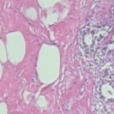 |
Guided Grad-CAM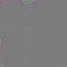 |
On natural images Grad-CAM localizes objects cleanly, but on H&E histopathology the attribution is diffuse and noisy — it does not isolate the diagnostically-relevant glands. This motivates an attention-based approach.
Attention-based Multiple Instance Learning (A-MIL)
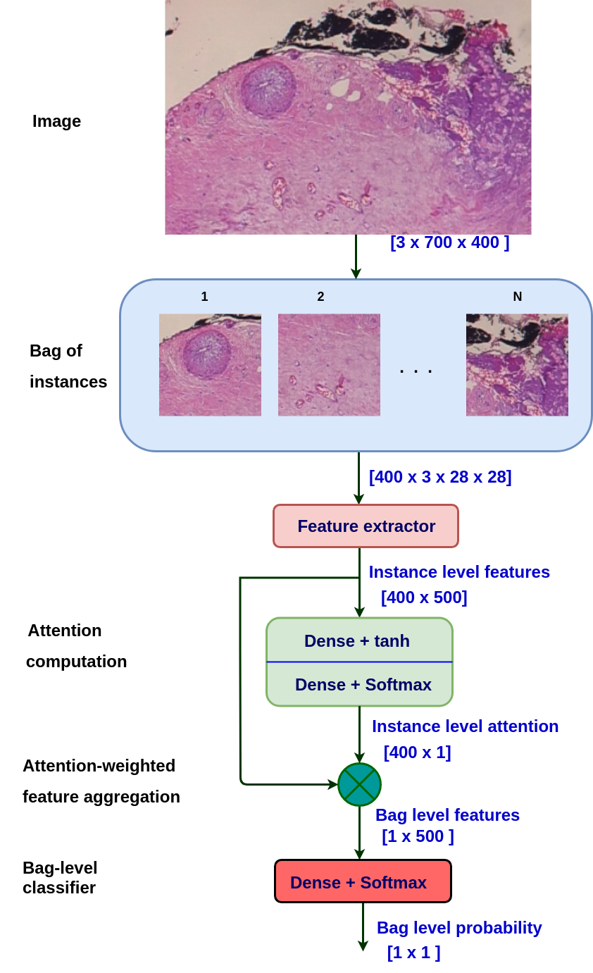
We crop each image into small patches to form a bag, extract per-patch (instance) features with a CNN, compute an attention weight for each patch, and aggregate the patches into a bag-level feature for classification (Benign / Malignant / Invasive):
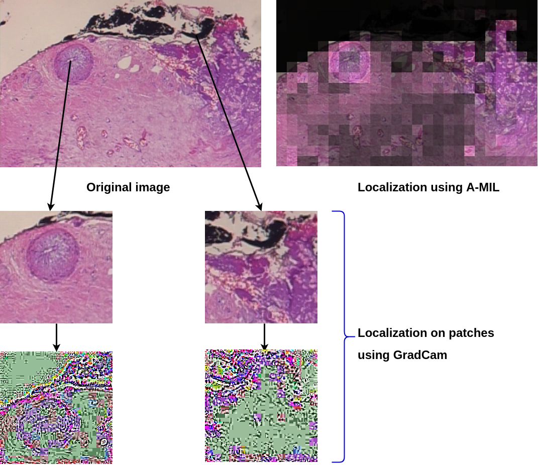
Multiplying each patch by its attention weight and stitching the patches back together reconstructs a localization map. A-MIL highlights the affected glands and ignores the background, while Grad-CAM on the same patches stays noisy:
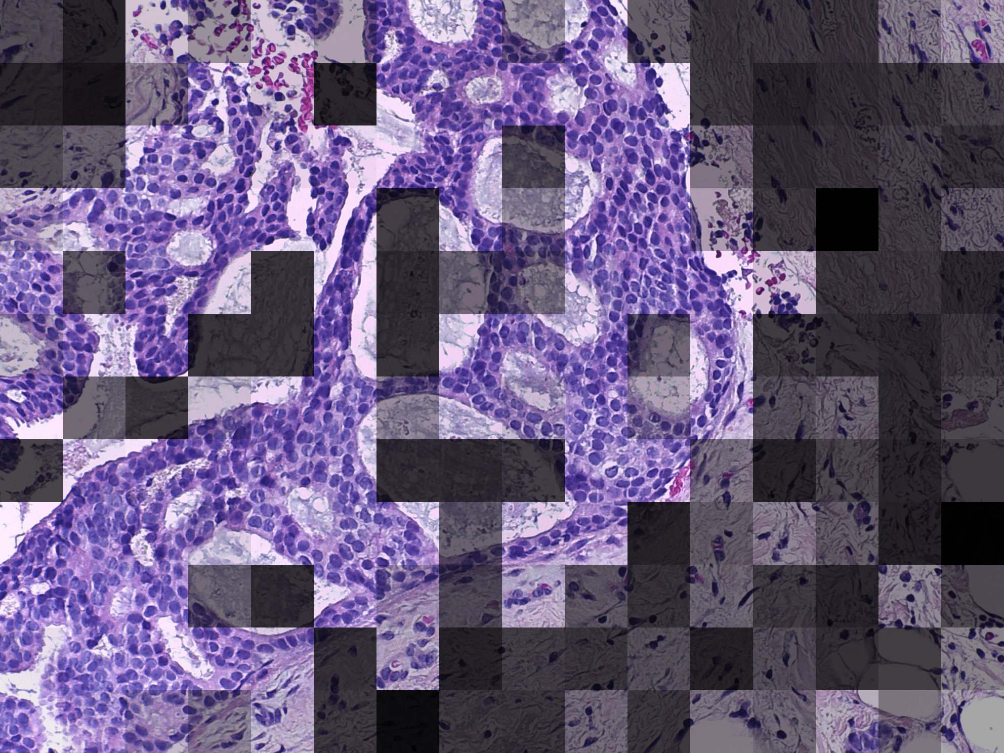
The same method applied to the BACH dataset (bright = attended informative regions, dark = ignored background):
Repository structure
| Path | Description | |------|-------------| | my_network/ | Custom CNN architecture — net.py | | ResNet/ | Transfer learning with a pretrained ResNet — resnet_pre.py | | VGG/ | Transfer learning with a pretrained VGG — vgg_pre.py | | AMILcodes/ | Attention-based MIL — model (amilmodel.py), patch/bag loader (patchdata.py), train + test (trainn_test.py) | | grad_cam/ | Grad-CAM / Guided Grad-CAM visualization toolkit | | AMILData/ | Expected data layout for A-MIL (readmedata_format.txt) |
How to run the code
Each model folder trains on the chosen magnification (zoom) level and saves the model, terminal logs, TensorBoard logs and a run summary into the corresponding zoom-level folder.
- Custom network:
cd my_network && python net.py - ResNet (pretrained):
cd ResNet && python resnet_pre.py - VGG (pretrained):
cd VGG && python vgg_pre.py - Attention MIL:
cd AMILcodes && python trainn_test.py— trains A-MIL, tests it, and saves the patch-stitched visualizations.
runforall_zoom.sh, which loops the corresponding script over all magnifications (40X, 100X, 200X, 400X):
bash runforall_zoom.shGrad-CAM visualization: put a test image in gradcam/inputs/, set its filename in gradcam/src/miscfunctions.py (around line 253), then run python gradcam/src/guidedgradcam.py. Results are written to gradcam/results/.
Dataset
BreakHis (used by the ResNet / VGG / custom-network models):
- Official source: Breast Cancer Histopathological Database (BreakHis), UFPR
- Also on Kaggle: BreakHis dataset
AMILData/readmedata_format.txt:
data_breakhis/
  40X/  100X/  200X/  400X/
    train/
      0/   (benign images)
      1/   (malignant images)
    test/
      0/   1/
Requirements
Note: this code was developed in 2019 on an older stack — PyTorch 0.4.1, torchvision 0.2.1, TensorFlow 1.13.1 (see requirement.txt). These pins no longer install on modern Python; to reproduce the original results, use a matching legacy environment (e.g. Python 3.6). Porting to a current PyTorch is straightforward but has not been re-validated here.
Links
Citation
@INPROCEEDINGS{9019916,
  author={Patil, Abhijeet and Tamboli, Dipesh and Meena, Sunil and Anand, Deepak and Sethi, Amit},
  booktitle={2019 IEEE International WIE Conference on Electrical and Computer Engineering (WIECON-ECE)},
  title={Breast Cancer Histopathology Image Classification and Localization using Multiple Instance Learning},
  year={2019},
  pages={1-4},
}
Plain text: A. Patil, D. Tamboli, S. Meena, D. Anand and A. Sethi, "Breast Cancer Histopathology Image Classification and Localization using Multiple Instance Learning," 2019 IEEE International WIE Conference on Electrical and Computer Engineering (WIECON-ECE), Bangalore, India, 2019, pp. 1-4.
License
Released under the MIT License.


